

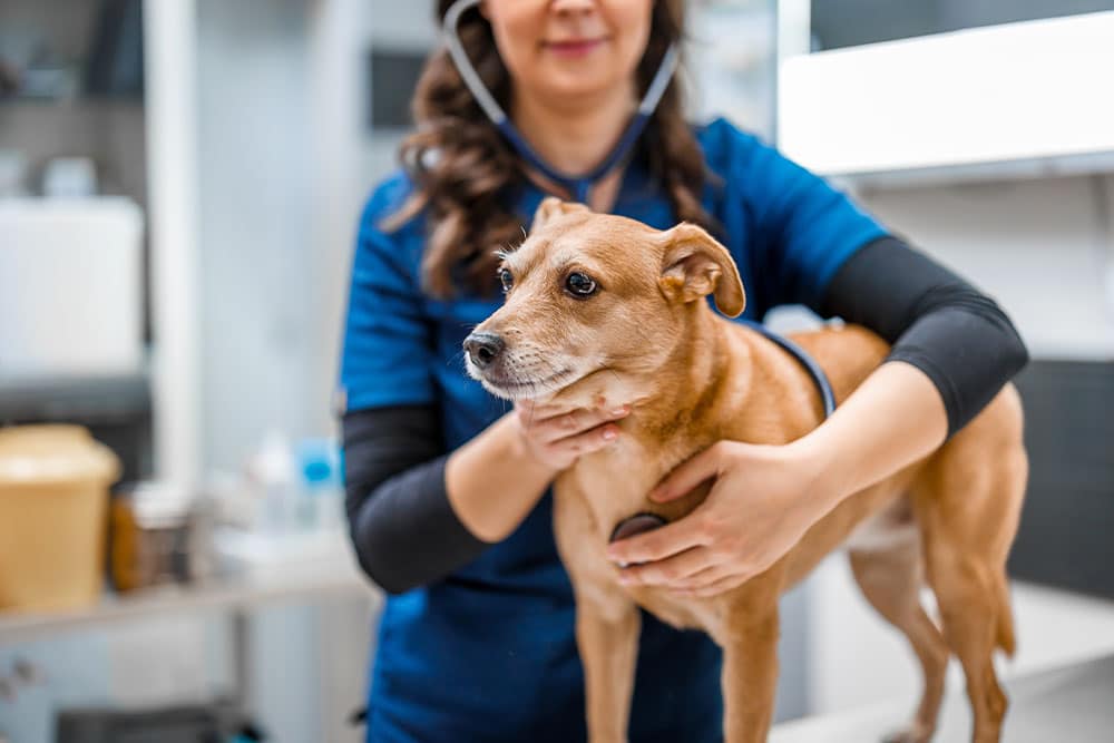
A veterinarian cannot reliably tell whether your pet’s lump is cancer just by feeling it, and neither can the most experienced specialist. A soft, squishy bump can turn out to be a mast cell tumor, and a firm, fixed one can turn out to be a harmless cyst. The only way to know what a mass actually is involves pulling cells or tissue from the lump and reading them under a microscope. The two routine options are a fine needle aspirate, which uses a small needle to collect a few cells, and a biopsy, which removes a larger piece of tissue. Which test fits depends on where the lump sits, how it behaves, and how much information the next decision needs.
Locally owned and rooted in Saint Petersburg, Bayview Animal Hospital starts every lump workup with a hands-on exam, in-house bloodwork, and a careful conversation about whether a fine needle aspirate or a biopsy is the right next step for your pet. We send samples to our reference lab for pathology and walk families through what an aspirate result or a biopsy result actually means for the decisions ahead. Our goal is to keep the workup measured, the timeline clear, and the next step matched to what the mass is doing. If you have noticed a new lump on your dog or cat, contact our team and we will take a careful look.
What Every Pet Family Should Remember
- No one, not even a board-certified specialist, can confirm whether a lump is cancer just by looking at it or feeling it, because benign and malignant masses can look and feel exactly the same.
- A fine needle aspirate is the quick, well-tolerated first step similar in feel to getting a vaccine, while a biopsy is a surgical procedure that removes actual tissue when the answer needs to include tumor grade, invasion, or margins.
- Some masses, like those on the spleen or in the intestines of cats, skip the aspirate entirely because a reliable answer requires whole tissue architecture.
- A benign result is good news but not the end of the story, since new masses can appear and some known ones change over time, which makes home monitoring worth the habit.
What Kinds of Lumps Do Dogs and Cats Get, and Why Can’t You Tell Them Apart by Looking?
Masses that look nearly identical on the surface can carry completely different prognoses, which is the whole reason sampling matters. A small wart that vanishes on its own and a cancer that spreads under the skin can both start as an unremarkable little lump. The types worth knowing differ a bit between the two species.
Which Skin Masses Are Common in Dogs?
Dogs grow a wide range of skin masses, and the common ones run the full span from harmless to serious. You cannot tell which is which by eye, which is exactly why each one gets sampled.
- Histiocytomas: These often show up as fast-growing red bumps on younger dogs. Most are harmless and shrink away on their own without treatment.
- Melanocytic tumors: These pigmented growths range from benign skin spots to aggressive melanomas, and where they sit on the body strongly influences how they behave.
- Cysts: Filled with fluid or other material, cysts are usually benign, but they can look identical to more concerning masses until they are sampled.
- Mast cell tumors: Mast cell tumors are notorious for masquerading as an ordinary bump while behaving aggressively underneath the skin, which is exactly why a benign-looking lump earns a closer look.
- Lipomas: Soft and movable, lipomas in dogs are benign fatty masses, though a lump that only feels like a lipoma still deserves confirmation rather than assumption.
Which Skin Masses Are Common in Cats?
Cats are experts at hiding illness, so feline tumors are often caught later than canine ones, which raises the stakes on any new lump. The masses below account for a large share of what we sample in cats, and feline mast cell tumors in particular can go unnoticed until they are advanced.
- Basal cell carcinoma: This often appears as a firm, sometimes pigmented mass. Its most common form is frequently benign, though it still needs sampling to confirm.
- Squamous cell carcinoma: Squamous cell carcinoma in cats often starts as a small crusty sore on the ears, nose, or eyelids that is easy to overlook until it has spread.
- Fibrosarcoma: Deeper and firm, fibrosarcoma in cats tends to invade local tissue aggressively, which makes finding it early especially valuable.
- Mast cell tumors: Typically raised, pink bumps on the head, neck, and trunk. Mast cell tumors in cats vary in how aggressively they behave, so they need a close look at cellular structure to know how to treat properly.
Besides a Lump, What Other Warning Signs Mean My Pet Should See a Vet?
Some of the most important red flags never show up as a mass at all, so a visible bump is only one of the things worth watching. Beyond the lump itself, the broader signs of cancer in pets include sudden weight loss, sores that will not heal, a shift in appetite, and swelling that changes in size or texture over time. Any of those, with or without a visible bump, is worth a conversation.
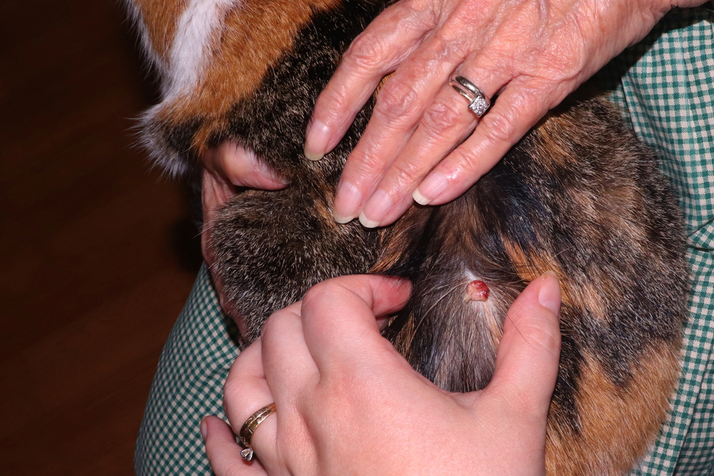
When a lump is what you have found, keep in mind that appearance is a poor predictor of what it actually is. A benign fatty deposit and an aggressive soft tissue sarcoma can both feel like a soft, freely movable lump under the skin, and the trick runs in reverse too, where a firm, worrisome-feeling nodule turns out to be a simple cyst. Feel and shape do not carry enough information to sort harmless from dangerous.
We know that is hard to sit with. You found something, you want to know what it is, and the honest answer is that we need to look at the cells before anyone can say for sure. Sampling turns a guess into a diagnosis, and that clarity is what every good next decision is built on.
What Is a Fine Needle Aspirate, and Why Is It Usually the First Test?
A fine needle aspirate uses a small needle to collect a few cells from your pet’s lump, and for most masses it is the right first test. It is quick, usually needs no sedation, and returns results within a few business days once a pathologist reads the cells, a process called cytology.
Most pets barely notice the aspirate itself, which feels similar to a routine injection. That is why fine needle aspiration is the sensible first step for most masses: a quick, well-tolerated procedure that draws out cells without fuss. For masses that sit deeper or in a spot that is hard to reach by feel, ultrasound might be used to guide the needle to the right place.
Cytology handles many tumor types beautifully and often confirms a lipoma, a mast cell tumor, or an infection outright. It has limits, though. Loose cells cannot always show how a tumor is organized, how aggressive it is, or whether it has invaded surrounding tissue. Not every tumor type releases its cells easily, so the test may come back showing nothing. When cytology comes back inconclusive or raises a question it cannot answer, the next step is usually a biopsy.
When Does My Pet Need a Biopsy Instead of a Needle Test?
A biopsy moves beyond individual cells to remove an actual piece of tissue which lets a pathologist read information that loose cells simply cannot show. The distinction between cytology versus biopsy comes down to whether the lab examines loose cells or an intact piece of tissue, and only tissue reveals a tumor’s grade, how far it has invaded, and whether surgical margins are clean.
That extra detail matters because the many types of cancer in pets behave so differently, and pinning down the exact tumor’s behavior with tissue is often what turns a vague diagnosis into a clear treatment plan. Biopsies come in a few forms. An incisional biopsy takes a wedge from a larger mass, an excisional biopsy removes the whole thing at once, and a needle-core biopsy pulls a thin cylinder of tissue. All of them require sedation or anesthesia, which is the main tradeoff against the quick, awake aspirate. Once the report is back, our veterinarians sit down with your family to walk through what the tissue actually showed.
| Feature | Fine Needle Aspirate | Biopsy |
| What is collected | A few loose cells | An intact piece of tissue |
| Sedation or anesthesia | Usually none | Required |
| Typical turnaround | A few business days | 5-10 days, depending on type |
| Tumor grade and margins | Not reliably shown | Assessed directly |
| Best used as | First step for most masses | When cytology is inconclusive or more detail is needed |
The Masses That Bypass the Needle Entirely
Some masses skip the aspirate entirely because a reliable answer depends on seeing how the tissue is built, not just what cells are present. In these situations the architecture of the tissue carries the diagnosis, and loose cells would either mislead or come back frustratingly unclear. Two good examples are hemangiosarcomas in dogs (a tumor of the spleen) and intestinal lymphoma in cats, though there are other cancers that also fall in this category.
Is a Splenic Mass Benign or Hemangiosarcoma?
A mass on the spleen is one of the hardest calls in veterinary medicine, since imaging and aspiration cannot reliably tell a benign nodule apart from hemangiosarcoma in dogs, and removing the spleen for tissue analysis is frequently the only way to know for certain. Aspirating a splenic mass also carries a real bleeding risk, since these masses originate from the cells of blood vessels.
Because of that, the recommendation is usually to remove the spleen and send it for histopathology, even before a definitive diagnosis is confirmed. It feels backward to operate before you have the answer, but doing so is often what produces it safely. Our surgical services cover splenectomy, and the removed tissue goes to the lab for the definitive read.
Is My Cat’s Chronic Vomiting IBD or Lymphoma?
Distinguishing inflammatory bowel disease from small-cell intestinal lymphoma in cats is one of the genuine puzzles in feline medicine, because the two look nearly identical everywhere except in the tissue itself. The vomiting, diarrhea, and slow weight loss of inflammatory bowel disease in cats look almost the same as small-cell intestinal lymphoma on exams, imaging, and bloodwork, and only a full-thickness intestinal biopsy can reliably separate the two.
That distinction is worth the effort because the treatment paths diverge sharply, and feline lymphoma calls for a very different plan than managing inflammatory bowel disease. A full-thickness biopsy gives the pathologist the depth of tissue needed to tell them apart with confidence.
Reading the Report: What Each Result Sets in Motion
Results land in one of three buckets, and each one points to a different next step rather than a single fixed path. Knowing the categories ahead of time takes some of the anxiety out of the waiting.
| Result | What it means | Typical next step |
| Benign | The mass is not cancer | Set up a monitoring plan and watch for change |
| Malignant | The mass is cancer | Open the treatment conversation: surgery, staging, oncology, or palliative care |
| Inconclusive | The sample could not give a clear answer | Move to further diagnostics, often a biopsy |
A benign result usually means we settle into a monitoring rhythm and get on with life. A malignant result opens a fuller conversation about surgery, staging to check whether cancer has spread, a referral to an oncologist, or palliative care focused on comfort, and we tailor that discussion to your pet and your family. When decisions get hard, our team offers compassionate support so no family faces them alone. An inconclusive result is not a dead end, just a signal to gather more information before deciding.
Keeping an Eye on a Lump That Turned Out Harmless
A benign result is reassuring, but it does not close the file, because new masses can develop alongside a known one and some lumps change over time. Home monitoring is how those shifts get caught early, and it takes only a minute now and then. The habit is simple: check the lump, and check the rest of your pet while you are at it.
A practical routine is to measure the mass with a soft tape or ruler, note its size and feel, and snap a phone photo every few weeks so you have a record to compare against. Call us if a benign lump grows quickly, changes shape or firmness, becomes painful, bleeds or scabs, or if a brand-new lump appears nearby. Even a confirmed lipoma sometimes earns removal down the road if it grows large enough to press on a joint or interfere with how your pet moves. When something shifts and you are not sure whether it matters, give us a call and we will help you decide whether it needs a look.
Can My Pet Be Screened for Cancer Before a Lump Ever Shows Up?
A newer category of cancer screening can flag disease before any lump forms by detecting genetic material that tumor cells shed into the bloodstream. A positive result is not a diagnosis on its own; it simply tells us to take a closer look with follow-up diagnostics.
These cancer screening blood tests, often called liquid biopsy screens, are especially useful for high-risk breeds. The lymphoma risk in Golden Retrievers is a well-known example of a breed predisposition where earlier detection genuinely changes options. Blood-based screening slots naturally into an annual senior exam and can prompt a closer look before anything is visible. For older pets and predisposed breeds, it is worth asking whether to schedule a wellness visit that includes this kind of screening.
Common Questions Pet Families Ask About Cytology and Biopsy
Does a fine needle aspirate hurt my pet?
Most pets tolerate an aspirate about as well as a routine vaccination. The needle is small, the procedure takes seconds, and sedation is usually unnecessary. Some pets flinch briefly, the same way anyone might at a quick pinch, but the discomfort passes almost immediately.
For masses in a sensitive or hard-to-reach spot, we may add light sedation or some local anesthetic to keep the experience calm and get a clean sample on the first try.
Can an aspirate spread cancer through my pet’s body?
For the vast majority of tumors, aspirating a mass does not cause it to spread, and the diagnostic benefit far outweighs the theoretical risk. This is a common worry, and a reasonable one to ask about. A small number of specific situations, such as certain splenic or bladder masses, call for a different approach precisely because of bleeding or spread concerns, which is one reason those masses often go straight to surgical biopsy rather than aspiration.
Why did my pet’s aspirate come back inconclusive?
Cytology relies on loose cells, and sometimes a mass simply does not release enough diagnostic cells, or the cells it releases cannot show how the tissue is organized. That does not mean anything was done wrong. Some tumor types, especially firm or fibrous ones, are notoriously hard to aspirate. An inconclusive result usually means the next step is a biopsy, which samples intact tissue and answers the questions loose cells could not.
How long do results take to come back?
Aspirate cytology results typically return within a few business days once the sample reaches our reference lab. Biopsy findings take longer, since the tissue has to be processed, sectioned, and stained before a pathologist reads it. We know the waiting is the hard part, and we will give you a realistic timeline when we send the sample so you are not left wondering.
Getting to a Clear Answer Sooner for Your Pet
Earlier testing simply gives you more room to act. The lump that turns out to be benign hands you relief, and the one that turns out to be malignant carries far more treatment options when it is caught early rather than late. Either way, knowing beats wondering.
If you have found a new lump or noticed a change in one you have been watching, our team can talk through whether an aspirate or a biopsy is the right next step with our in-house diagnostics and surgical care. When you are ready, book a visit and we will take a careful, unhurried look.




Leave A Comment